Contact
Fields of application & application examples
Revolutionary ultrasound diagnostics in dentistry
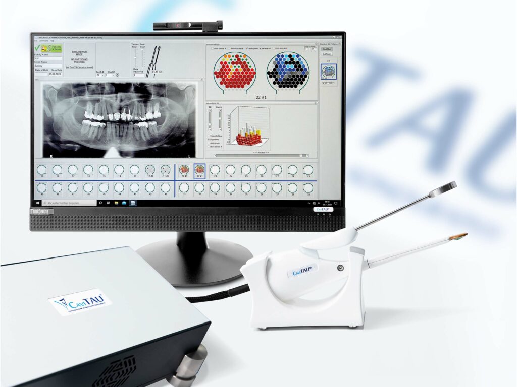
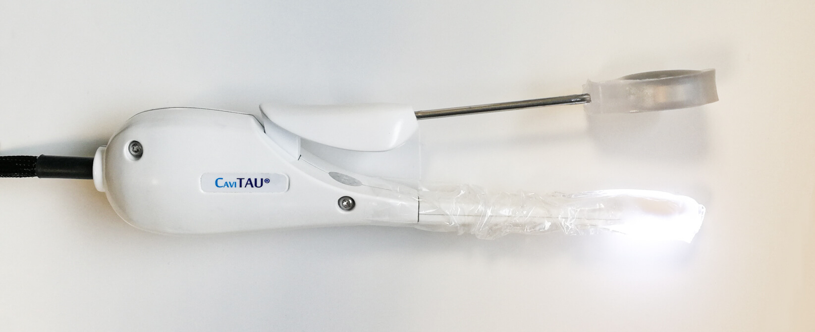
Alveolar Transmission Ultrasound or Trans Alveolar Ultrasound Diagnostics, which is being implemented in CaviTAU®, is an imaging procedure in dentistry. This ultrasound diagnostic is necessary for the early detection and treatment of jawbone with low bone density, such as in areas of ischemic osteonecrosis.
In terms of the legally prescribed duty of care, CaviTAU® can be used as a non-invasive and cost-effective adjuvant for medical indication in the event of the following procedures:

Suspicion of jawbone necrosis with chronic facial pain

Suspicion of jawbone necrosis with immunological and tumour diseases

To protect the resilient bone during implantations to ensure the success of dental implants.
Application examples:
CaviTAU® detects so-called “bone marrow defects” in the jawbone as RANTES/CCL5 sources in the context of focal areas of inflammation.
By developing CaviTAU®, we are thus able to detect pre-inflammatory and pre-cancerous niches suffering from “silent inflammation” in the jawbone
CaviTAU® reveals reduced mineral density and focal areas of inflammation around implants.
By developing CaviTAU®, we are thus able to detect unexplained local, systemic complaints in implant wearers
CaviTAU® reveals reduced mineral density and focal areas of inflammation around teeth that have undergone root canal treatment.
By developing CaviTAU®, we are thus able to detect unexplained local, systemic complaints in patients who have undergone root canal treatment
CaviTAU® displays perfectly healed, stress-free jawbones for easy implant insertion.
By developing CaviTAU®, we are able to achieve higher rates of success for implants without “inexplicable” implant losses
CaviTAU® can also play an important role in the context of clinical research into factors that contribute to different forms of systemic diseases.
Why is it so difficult to diagnose fatty-degenerative osteolysis / osteonecrosis in the jawbone (FDOJ)?
The problem: On X-ray images, the bone dissolution of FDOJ areas cannot be determined with certainty. Suspected sites in the jaw can be confirmed or excluded with CaviTAU®. From the sum total of anamnesis, X-ray image OPG, X-ray image CBCT, lymphatic-palpatory findings and the CaviTAU® bone density measurement, you can finally get an idea of whether surgical intervention on the jaw is necessary.
- [Al-Nawas B et al, Dental Implantation: Ulrasound Transmission Velocity to Evaluate Critical Bone Quality – An Animal Model. Ultraschall in Me/European Journal of Ultrasound 2008; 29:302-307.]
- [Lechner, J. Validation of dental X-ray by cytokine RANTES – comparison of X-ray findings with cytokine overexpression in jawbone. Clinical, Cosmetic and Investigational Dentistry 2014:6 71–79]

Why is CaviTAU® necessary in dentistry and medicine?
In the medical field in general, pulse-echo ultrasonography is widely used for imaging all kinds of soft tissue. In principle, images from structures inside the body are produced by analyzing the reflection of ultrasound waves. However, this method is not suitable to yield helpful information on the status of the jawbone because of the almost total reflection of ultrasound at the bone/soft tissue interphase. Particularly the cancellous part of the jawbone cannot be examined with commonly used ultrasound equipment. Thus, up to now, ultrasound was of minimal use in dental medicine.
The status of the cancellous bone of the jaw can be of great clinical importance. Jerry Bouquot has provided anatomical evidence that the cancellous bone can be largely degenerated, a phenomenonhe calls inter alia “ischemic osteonecrosis leading to cavitational lesions”. He relates osteonecrosis of the jawbone to neuralgic pain and defines a disease called “neuralgia inducing cavitational osteonecrosis (NICO)”
Dr. Johann Lechner investigated in-depth the tissue in such lesions, which appears as a clump of fat inside an intact cortical bone. During surgery, the material is simply spooned out. This tissue is in an ischemic, fatty degenerative state. Dr. Lechner, therefore, defines the observed changes as “fatty-degenerative osteolysis/osteonecrosis of the jawbone (FDOJ)”. He showed that the clumps of fat found in the jawbone are biochemically exceedingly active, producing certain cytokines in high amounts, namely RANTES (CCL-5) and FGF-2, PDGF, and MCP-1. The level of these cytokines is also elevated in several systemic diseases such as cancer, dementia, multiple sclerosis, or arthritis. Strong evidence suggests that eliminating such tissue surgically supports clinical improvement. Furthermore, the development and the persistence of various systemic diseases can be related to the fatty-degenerative osteolysis of the jawbone (FDOJ).
Despite, in most cases, the local effect of neuralgic pain (NICO) is missing.
Furthermore, in a recent publication, it was made plausible that NICO and FDOJ, as well as the so-called “aseptic ischemic osteonecrosis in the jawbone” (AIOJ), all, describe the same pathological condition of the jawbone. This condition is listed under Code M87.0 in the International Statistical Classification of Diseases and Related Health Problems, tenth revision (ICD-10).
Additionally, the status of the cancellous part of the jawbone is of great importance for dental implants and for the success of implantology, according to earlier publications of Bilal AI-Nawas.
Hence, serious health risks can be associated with fatty-degenerative osteolysis of the jawbone. However, the major problem is that a jawbone with fatty-degenerative osteolysis appears without abnormal findings in X-ray examination. This holds true even if the cancellous bone is in a primarily degenerated status, as it shows only fatty tissue instead of the substantia spongiosa of healthy cancellous bone (FDOJ). Being virtually undetectable by any kind of X-ray examination, the occurrence and the phenomena of AIOJ, FDOJ, and NICO remain widely unknown and even are disputed or denied.
To overcome the aforementioned problem, a different approach was needed. Instead of X-ray or other established medical examination methods, the use of Through-Transmission Alveolar Ultrasonography (TAU) was created.


